
In the rapidly evolving landscape of artificial intelligence, the "bigger is better" mantra has largely dominated the narrative, fueled by the success of massive transformer models like GPT and DINO. However, in the high-stakes domain of medical imaging, a new breakthrough suggests that strategic efficiency and domain expertise may be more valuable than sheer computational scale. A research team led by Pedro M. Gordaliza, Jaume Banus, and Benoît Gérin has demonstrated that compact, specialized models can not only compete with but significantly outperform their larger counterparts in the complex task of 3D brain MRI analysis.
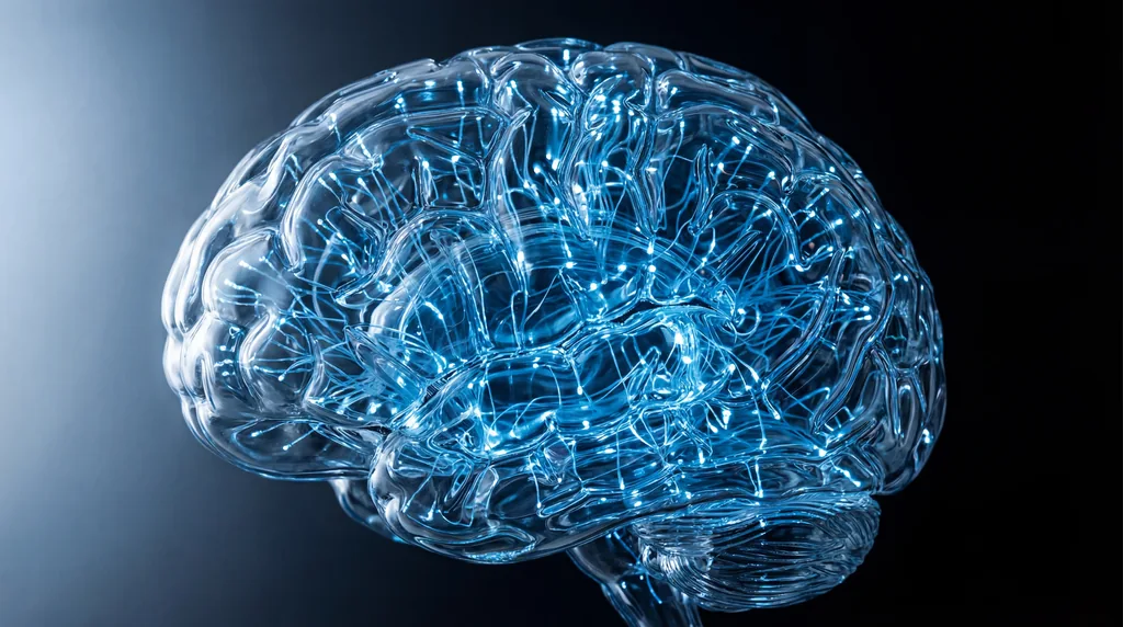
The Rise of Brain MRI Foundation Models
Foundation models (FM) represent a paradigm shift in artificial intelligence. Unlike traditional models trained for a single specific task, foundation models are pre-trained on vast, unlabeled datasets using self-supervised learning (SSL), allowing them to be fine-tuned for a wide variety of downstream applications with minimal labeled data. While these models have revolutionized natural language processing and 2D computer vision, their application to 3D medical imaging—specifically neuroimaging—has remained a formidable challenge. The brain's anatomical complexity, coupled with the high-dimensional nature of volumetric MRI data and the variability in acquisition protocols, creates a unique bottleneck for standard AI architectures.
To address these barriers, the medical imaging community established two landmark competitions at the MICCAI 2025 conference: the Self-Supervised Learning for 3D Medical Imaging Challenge (SSL3D) and the Foundation Model Challenge for Brain MRI (FOMO25). These contests served as the first rigorous, standardized benchmarks for evaluating how well foundation models can generalize across heterogeneous clinical datasets. The SSL3D challenge alone compiled an unprecedented dataset of over 114,000 3D volumes from 34,191 subjects, spanning 800 different datasets. It was within this competitive arena that the research team, representing institutions including the Lausanne University Hospital (CHUV), the University of Lausanne (UNIL), and the CIBM Center for Biomedical Imaging, secured first-place rankings using a surprisingly lean approach.
Small AI vs. Massive Transformers
One of the most striking findings from the researchers' success is the continued dominance of Convolutional Neural Networks (CNNs), specifically the U-Net architecture, over the currently fashionable Transformer-based models. In the FOMO25 and SSL3D challenges, none of the transformer-based submissions managed to match the performance of the winning CNN method. This disparity highlights a critical technical limitation: Transformers, while powerful in 2D or text-based tasks, suffer from quadratic complexity when processing the massive token counts generated by 3D volumetric tokenization. This creates a computational bottleneck that limits the spatial resolution and context these models can effectively manage.
The research team’s model achieved its top-tier performance while being approximately 10 times smaller than competing transformer-based approaches, such as the ViT-L DINOv2 3D. While larger models often boast hundreds of millions of parameters, the winning CNN-based architecture utilized only 20 million. Despite this smaller footprint, the team reported a 2.5% higher average Dice score for segmentation tasks and an 8% increase in accuracy for classification tasks compared to transformer-based rivals. This suggests that the "bitter lesson" of AI—that general methods eventually win through scale—may not yet apply to the intricate, resource-constrained world of 3D medical imaging.
The Power of Domain Knowledge
The secret to the team’s success lay in the integration of anatomical priors and neuroimaging domain knowledge into the model's architecture. Instead of treating the 3D volumes as generic data points, Gordaliza, Banus, and Gérin designed their system to disentangle subject-invariant anatomical structures from contrast-specific pathological features. By forcing the model to recognize that certain anatomical features remain consistent across different MRI contrasts (like T1-weighted or T2-weighted images) and timepoints, they provided the neural network with an "inductive bias" that prevents it from learning spurious correlations or taking computational shortcuts.
For the SSL3D challenge, the researchers partitioned learned representations into two distinct components: one constrained to match anatomical segmentations across all images of a single subject, and another optimized to detect pathology. In the FOMO25 track, they implemented a cross-contrast reconstruction objective, swapping representations between different scans of the same subject during pre-training. This domain-specific guidance allowed the model to focus on what truly matters in a clinical context—the underlying biological reality—rather than getting lost in the noise of varying scanner manufacturers or acquisition settings.
Speed and Efficiency Benchmarks
The practical implications of this research extend beyond accuracy scores; the gains in efficiency are equally transformative. The team reported that their models trained one to two orders of magnitude faster than transformer alternatives. In the FOMO25 challenge, the CNN model required fewer than 36 GPU-hours for pre-training, compared to the 100 to 1,000 hours required by larger transformer models. This reduction in training time not only accelerates the pace of research but also significantly lowers the carbon footprint associated with developing high-end medical AI.
Furthermore, this "efficiency-first" approach democratizes access to foundation models. While massive 7-billion-parameter models like DINOv3 require industrial-scale computing clusters, the team’s 20-million-parameter model can be trained and fine-tuned on hardware accessible to smaller research institutions and hospitals. This accessibility is vital for the clinical deployment of AI, where models must often be adapted to local hardware constraints and specific patient populations without the need for massive server farms.
Open Science and Future Implications
In a commitment to open science, the researchers have made their winning models and code available via GitHub at jbanusco/BrainFM4Challenges. By sharing these tools, they aim to provide a robust starting point for other researchers to build upon, potentially accelerating the development of what some call "Artificial General Intelligence (AGI) for healthcare." The team’s work underscores a growing realization in the field: the path to universal medical AI may not be paved with more parameters, but with smarter, more principled exploitation of existing medical knowledge.
Looking ahead, the success of these compact models raises important questions about the future trajectory of AI in medicine. While it remains to be seen if transformers will eventually overcome their current limitations with even larger datasets or more efficient attention mechanisms, the lessons from MICCAI 2025 are clear. For now, the most effective way to analyze the human brain is to build AI that "understands" the brain's structure from the ground up. As the field moves toward more generalizable models, the integration of longitudinal trajectories, complementary contrasts, and anatomical priors will likely remain the gold standard for clinical AI development.

Comments
No comments yet. Be the first!