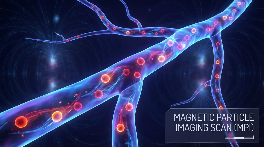

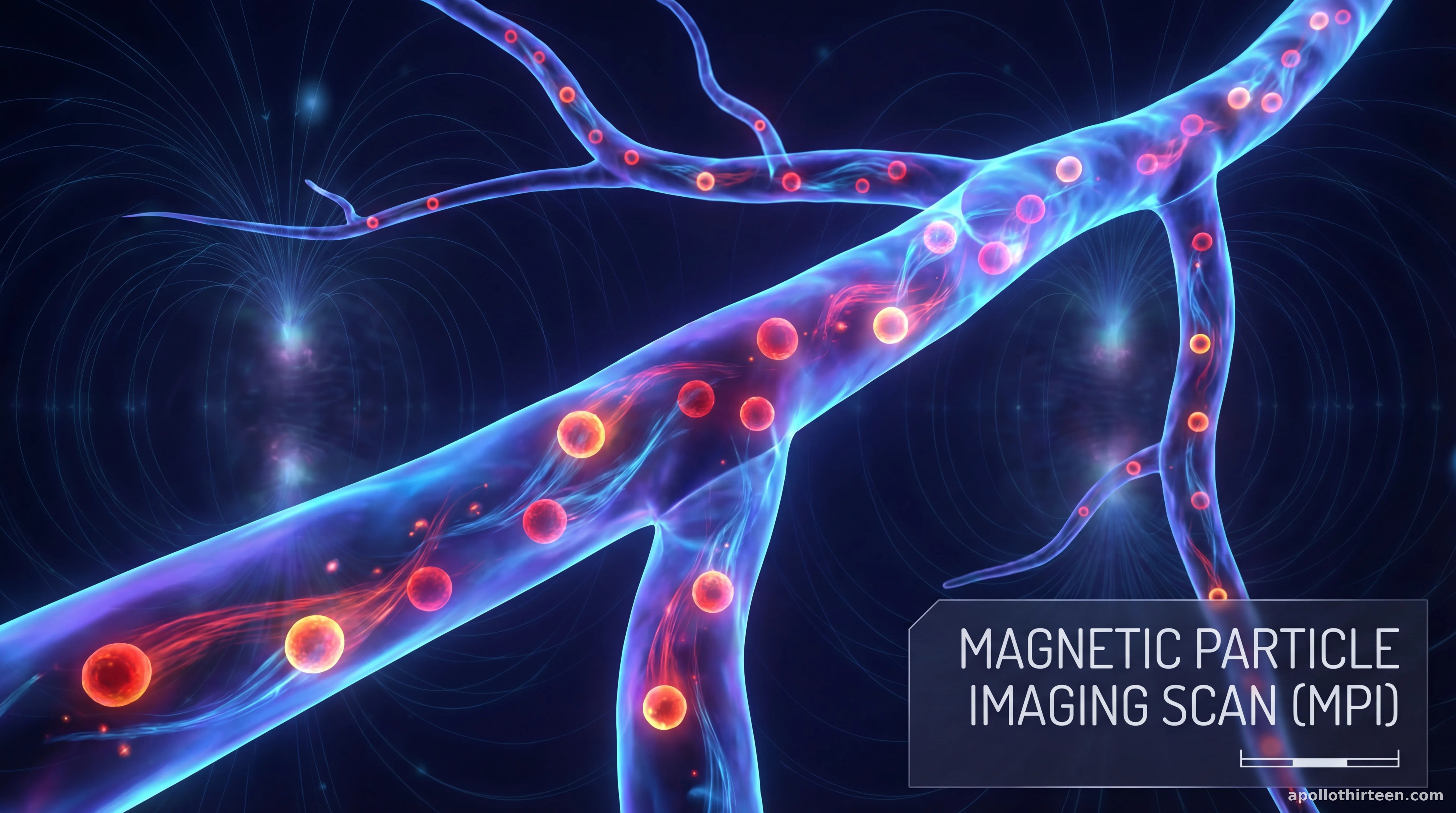
How Does Magnetic Particle Imaging Differ from MRI?
Magnetic Particle Imaging (MPI) differs from traditional MRI by directly detecting the magnetization of injected superparamagnetic iron-oxide nanoparticles rather than measuring the nuclear magnetic resonance signals from hydrogen atoms in water protons. This fundamental fusion of high-sensitivity tracer detection and rapid signal processing allows for superior contrast and the total elimination of background signals from surrounding biological tissues.
While Magnetic Resonance Imaging (MRI) is an indispensable tool in modern medicine, its reliance on the body’s internal water molecules often results in complex background noise that can obscure delicate vascular structures. In contrast, MPI functions as a purely tracer-based modality. By utilizing a "selection field" that creates a field-free point (FFP), the human-scale MPI scanner can pinpoint the exact location of nanoparticles with millisecond temporal resolution. This distinction is critical for clinicians who require precise, real-time data without the interference of bone or dense soft tissue, which often complicates traditional imaging interpretations.
The research led by Patrick Vogel, Thomas Kampf, and Martin A. Rückert marks the first time this technology has successfully transitioned from preclinical models to a living human subject. Their study highlights how MPI's unique physics allows for a zero tissue background, providing a high-contrast map of the circulatory system. This breakthrough suggests that MPI could soon complement or even replace certain diagnostic protocols that currently rely on the bulky and slower processes inherent to proton-based resonance imaging.
Is MPI Safe for Human Use with Iron-Oxide Nanoparticles?
Magnetic Particle Imaging is safe for human use because it utilizes biocompatible iron-oxide nanoparticles and operates entirely without ionizing radiation, unlike X-ray or CT scans. This technological fusion of magnetic safety and tracer efficiency makes it an ideal alternative for patients who cannot tolerate traditional contrast agents like iodine or gadolinium, particularly those with chronic kidney disease.
The safety profile of iron-oxide nanoparticles, such as the clinically approved Ferucarbotran used in this trial, is a significant advantage over existing methodologies. In traditional Digital Subtraction Angiography (DSA) and Computed Tomography (CT), patients are exposed to ionizing radiation and nephrotoxic contrast media that can lead to long-term health complications. Because MPI tracers are metabolized naturally by the liver and integrated into the body's iron stores, the risk of renal toxicity is virtually eliminated, making the procedure repeatable for frequent monitoring of chronic conditions.
Furthermore, the Vogel et al. study demonstrated that the in-vivo application of MPI did not produce adverse effects during the visualization of the upper extremity. The researchers utilized a human-scale scanner designed to maintain safety limits regarding peripheral nerve stimulation (PNS) and specific absorption rates (SAR). By successfully performing the first human angiography with a clinically approved tracer, the team has validated that the magnetic field strengths required for high-resolution human imaging remain well within established safety thresholds for medical devices.
What are the Advantages of MPI for Human Imaging?
The primary advantages of MPI include radiation-free imaging, tenfold higher sensitivity than functional MRI, and the ability to track biological processes in real-time at 2 frames per second. This fusion of speed and safety allows for persistent monitoring, as MPI tracers remain detectable for days or weeks, whereas PET tracers decay within hours.
In addition to safety, the temporal resolution of MPI offers a transformative leap for dynamic vascular assessment. During the first human trial, the system captured:
- Venous perfusion of the upper extremity in real-time.
- Complex branching and inflow patterns within deep and superficial veins.
- The mechanics of valve filling and clearance dynamics.
- A direct comparison with X-ray digital subtraction angiography (DSA), confirming MPI's accuracy.
The Mechanics of the First Human MPI Angiography
The successful execution of the first human trial required a sophisticated human-scale MPI scanner capable of generating the precise magnetic gradients necessary for large-bore imaging. Previous iterations of MPI were limited to small-animal models due to the immense power and cooling requirements of the magnets. However, the system designed by the research team overcame these engineering hurdles, allowing a human arm to be positioned within the imaging volume to capture angiographic data with unprecedented clarity.
During the procedure, the researchers administered Ferucarbotran, an iron-oxide-based contrast agent, into the subject's venous system. The scanner then tracked the movement of these particles as they flowed through the vasculature of the forearm. Unlike traditional methods that take a "snapshot," the MPI system recorded the distribution of nanoparticles at a rate of 2 frames per second. This high-speed data acquisition allowed the team to observe the physiological movement of blood, including the opening and closing of venous valves, which is often difficult to visualize using conventional static imaging.
Comparative Analysis: MPI vs. Digital Subtraction Angiography
To validate the findings, the researchers performed X-ray digital subtraction angiography (DSA) under identical procedural conditions, as it remains the clinical gold standard for vascular imaging. The DSA results provided a benchmark for the MPI data, confirming that the new modality could accurately identify major superficial and deep veins. Remarkably, the MPI images provided similar structural detail but without the "ghosting" effects or bone interference common in X-ray-based techniques.
The comparison revealed that MPI possesses a unique "hot spot" imaging characteristic. Because there is no signal from the surrounding muscle, bone, or fat, the resulting image is a pure representation of the tracer distribution. This leads to an exceptionally high signal-to-noise ratio (SNR). Scientists noted that while DSA requires complex post-processing to "subtract" the bone and background, MPI produces a clean vascular map natively, simplifying the diagnostic workflow and reducing the potential for human error in image interpretation.
Clinical Implications for Chronic Vascular Care
The transition of MPI from a preclinical curiosity to a clinically translatable modality has profound implications for long-term patient care. Patients suffering from chronic conditions such as peripheral artery disease, varicose veins, or deep vein thrombosis often require multiple imaging sessions over several years. The cumulative ionizing radiation from these sessions is a known risk factor for secondary malignancies; MPI offers a pathway to mitigate this risk entirely.
Furthermore, the scalability of MPI hardware suggests that future iterations could accommodate full-body scans. This would allow for radiation-free monitoring of cardiovascular health, organ perfusion, and even the tracking of stem cells or immune cells labeled with iron particles. The ability to monitor cell-based therapies in real-time could revolutionize oncology and regenerative medicine, providing a window into how the body responds to treatment at a molecular level without compromising the patient's systemic health.
The Future of Magnetic Particle Imaging
Moving forward, the research team aims to refine the human-scale hardware to improve spatial resolution further and expand the field of view. While the first trial focused on the upper extremity, the next phase of development will likely target cerebral blood flow and cardiac imaging. These applications require even faster acquisition speeds and more powerful magnetic gradients, but the success of this first in-vivo trial provides the necessary proof-of-concept to secure further investment and regulatory interest.
In conclusion, the work of Vogel, Kampf, and Rückert establishes a landmark in medical physics. By proving that Magnetic Particle Imaging can be safely and effectively applied to humans, they have opened the door to a new era of nanomedicine. As hardware continues to evolve and new specialized nanoparticles are developed, MPI is poised to become a cornerstone of diagnostic radiology, offering a safer, faster, and more precise look inside the human body than ever before.


Comments
No comments yet. Be the first!